
 According to a study, published in the December issue of Cell Reports, microglia, the brain’s immune cells responsible for its response to infection or injury, may also regulate the brain’s response to diet, and, thus play a role in obesity.
Fatty acids (FAs) serve as energy substrates and as signals controlling metabolic processes.
The brain’s mediobasal hypothalamus (MBH) is able to sense fatty acids (FAs) and this process is involved in the control of food intake, thermogenesis and metabolism. In addition, Diet-induced obesity also produces metabolic inflammation in the MBH and work targeting TLR4, tumor necrosis factor α (TNF-α), NF-κB, and NLRP3.
Chronic consumption of saturated fat leads to inflammation in the adipose tissue, liver, and skeletal muscle, and in the periphery, the hallmark of this over-nutrition or ‘metabolic inflammation’ is the extensive accumulation of macrophages.
This type of inflammation also occurs in the hypothalamus, with accumulation of astrocytes and microglia, the brain’s analogs of macrophages, but it is unclear what orchestrates this process.
Metabolic inflammation in the MBH is marked by accumulation of astrocytes and microglia, the CNS analogs of macrophages. Yet, microglial responses to FAs are not understood, and tools to manipulate hypothalamic microglia have been lacking.
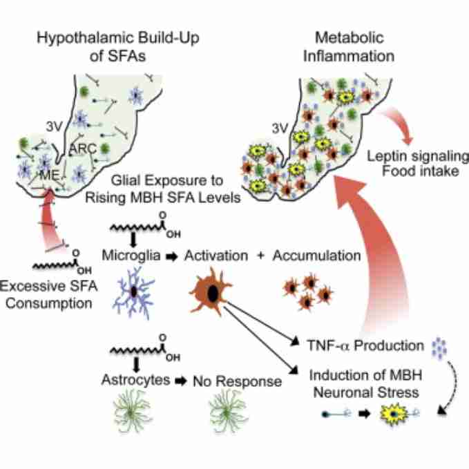
In the Cell Reports study a research team from UC San Francisco identifies hypothalamic microglia as sensors of saturated fat that are activated by rising levels of FAs, and that control the intensity of a highly localized form of inflammation in cases of high dietary intake. Microglial cells appear to mediate key changes in hypothalamic function that occur in response to consuming excess saturated fat, which include the reduction of the hypothalamic responsiveness to leptin, thus impacting food intake.
According to the authors, their study demonstrates that the transit of dietary SFAs into the brain is fast enough to stimulate the rapid inflammatory activation and proliferation of microglia previously reported.
Importantly, the SFA consumption induced microglial activation in the MBH despite controlling for total fat and caloric intake, and without increasing body weight. And as discussed by the authors, these data support their in vitro data, indicating that dietary SFAs directly stimulate M1 activation of MBH microglia, whereas peripheral tissue inflammation may depend, at least in part, on the presence of obesity.
Additionally, enteric SFAs induced MBH inflammation despite not increasing circulating levels of inflammatory cytokines, indicating that SFA-induced MBH inflammation is not a by-product of systemic inflammation.
Moreover, the investigators, by racking CCR2+ monocytes, were able to demonstrate that accumulation of MBH microglia in mice receiving SFA gavage was not due to infiltrating monocytes differentiating into microglia-like cells. Instead, the authors found that this accumulation is due to local proliferation in the MBH, a capacity that was inducible by depleting hypothalamic microglia using Lip-CLO or DT.
The study also provided some evidence indicating that whereas microglia sense SFA levels and transduce this into an inflammatory response, other cell types in the MBH may respond to monounsaturates.
According to Suneil Koliwad, the senior author of this study, “microglial activation in the brain may be a part of a normal physiological process to remodel brain function in response to changes in the composition of food intake”. The study also suggests that targeting hypothalamic microglia may be a promising way to mitigate diet-induced metabolic dysfunction.
Source: Cell Reports, 9, 1–15, December 24, 2014 DOI: http://dx.doi.org/10.1016/j.celrep.2014.11.018
Updates
A 2017 study by Yuanqing Gao et al. found that consumption of high-carbohydrate high-fat (HCHF) diets, but not of low-carbohydrate, high-fat (LCHF) diets, increases microgliosis as well as the presence of N(ε)-(Carboxymethyl)-Lysine (CML), a major advanced glycation end-products (AGEs), in POMC and NPY neurons of the arcuate nucleus. Neuron-secreted CML binds to both receptor (RAGE) and/or the activated leukocyte cell-adhesion molecule (ALCAM), which are expressed on endothelial cells, microglia, and pericytes.
The authors concluded that the combined overconsumption of fat and sugar, but not the overconsumption of fat per se, leads to excessive CML production in hypothalamic neurons, which, in turn, stimulates hypothalamic inflammatory responses such as microgliosis and eventually leads to neuronal dysfunction in the control of energy metabolism.
Calorie restriction is one of the major approaches to combat obesity and its associated metabolic disorders. According to the authors the evidence they provide indicates that restricting dietary fat is not the only factor that needs to be considered for body weight reduction in obese individuals and that dietary carbohydrates might substantially gate the efficiency of calorie restriction for body weight reduction, via a hypothalamic mechanism.
Another 2017 study by Martin Valdearcos et al. identified microglia as sensors activated by rising levels of dietary saturated fatty acids (SFAs) in the mediobasal hypothalamus (MBH) that control the intensity of a highly localized form of inflammation. In this setting, microglia mediate the stress-inducing effects of dietary SFAs on neurons residing in the arcuate nucleus (ARC) and reduce their responsiveness to leptin, thus impacting food intake.
 Highlights
– Saturated fats build up specifically in the hypothalamus when consumed in excess.
– Hypothalamic microglia directly and specifically sense saturated fatty acids.
-Microglia orchestrate hypothalamic inflammation due to excess saturated fat intake.
-Microglia dictate the impact of high dietary saturated fat on hypothalamic function.
Background: Diets rich in saturated fat produce inflammation, gliosis, and neuronal stress in the mediobasal hypothalamus (MBH). Metabolic inflammation in the MBH is marked by accumulation of astrocytes and microglia, the CNS analogs of macrophages. Based on the role of macrophages in metabolic inflammation, it is intriguing to consider targeting microglia to control CNS metabolic inflammation.
Metabolic inflammation in the MBH occurs more acutely in response to the steady consumption of excess saturated fat than in peripheral tissues, where it occurs in conjunction with obesity. There is interest in mitigating MBH inflammation due to this temporal primacy and because it is linked to metabolic dysregulation.
A 2021 review by Natália Ferreira Mendesr et al. explored microglial heterogeneity in the hypothalamus and their crosstalk with astrocytes under high fat diet–induced inflammation. The authors presented novel currently available ex vivo and in vivo experimental models that can be useful when designing a new research project in this field of study. They examined the transcriptomic data already published to identify how the hypothalamic microglial signature changes upon short-term and prolonged high-fat feeding.
Background: Microglia were first recognized as macrophage-like cells from the CNS a century ago, but for a long time their complexity was unknown. Luckily, the development of assorted transcriptomic tools has boosted the knowledge about these cells in recent years. Currently, it is well known that the hypothalamus presents several microglial subsets that can be identified by their hallmarks: Iba1, Cx3Cr1, Tmem119, P2ry12, Trem2, Hexb, and Csfr1, among many other.
In fact, microglia play a pivotal role in different stages of the hypothalamic inflammatory process, but how each microglial subtype reacts to SFAs from the diet, communicates with other cells, or even leads to the recruitment of peripheral myeloid cells remains to be explored.
Related stories you may like:
The Reward System and How it is Linked to Stress and the Epidemic of Obesity
Curvy Body, Newborns’ Brains and Breastfeeding: Why Do Women Need Fat?