
The immune- and nervous systems have historically been seen as two different entities but ongoing research is continuously unraveling the undeniable connection between the two. How are immunological organs such as lymph nodes innervated by the nervous system? What does a neuro-immune synapse look like and what other structures remain unknown in the interdisciplinary field of neuro-immunology?
These and more questions inspire our group daily. That’s why we use a multidisciplinary research approach working in continuous exchanges with other institutions like University Medical Center Hamburg-Eppendorf (UKE), DESY/ Helmholtz-Center Hereon, Bernhard Nocht Institute for Tropical Medicine (BNITM), The Imaging Center Essen (IMCES) and the Charité University of Medicine Berlin as well as exchange within our group.
Our research focus is the bi-directional communication between the nervous- and the immune system within the field of neuroimmunology. We are particularly interested in, how the autonomic but also the somatic part of the nervous system can influence physiological as pathophysiological immune responses. Additionally, we are eager to uncover how the immune system interacts with the central nervous system and ultimately shapes its cognitive functions. We mainly work on murine tissues in order to describe these interfaces.
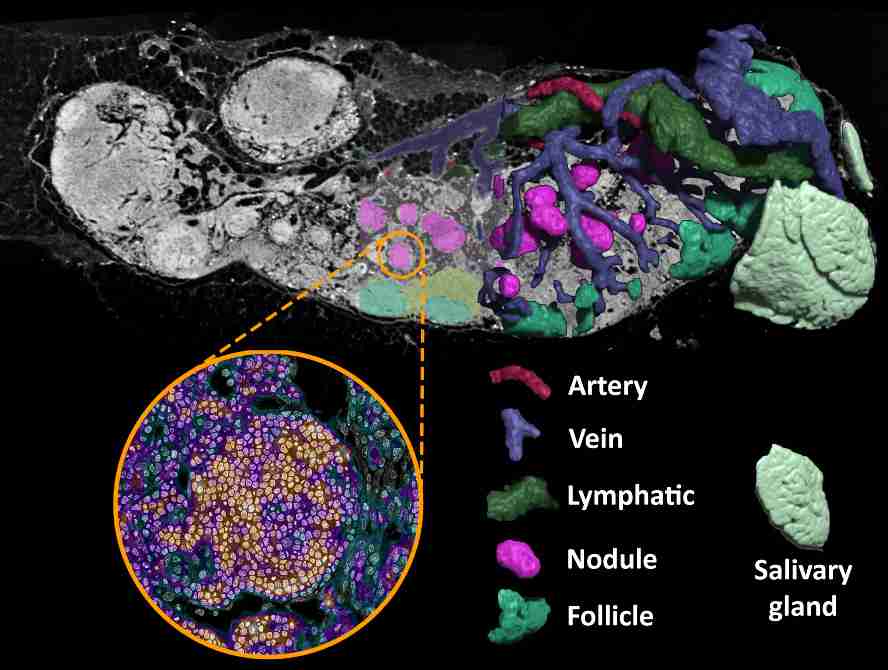
 Figure 1: 3D reconstruction of the structural composition of the lymphatic vessels, blood vessels, and cell areas of a mandibular mouse lymph node through synchrotron radiation-based μCT. It shows the spatial and structural separation of newly discovered B-cell nodules (magenta) with the already known B-cell follicles (mint green). These agglumerates of quiescent B cells can be found exclusively in mucosa-draining lymph nodes and their role in mucosal tolerance is currently under investigation. Correlative electron microscopy imaging reveals the ultrastructure of the densely packed B-cell nodules and their surroundings through loose tissue of the medulla and medullary sinuses. Image: INI Research/ Paul Schütz.
Current projects:
- Nodule quantification in three-dimensional lymph nodes using BABB-clearing: Paul Schütz and Merle Bannick are currently working on our nodules project specifically the quantification of nodules in different mouse models (germfree vs. SPF vs. wildlings) to see how the nodule number varies in different microbial environments. We want to investigate the nodules role in mucosal tolerance to find out more about their immunological role.
- Classification of sensory neuron subtypes and innervation pattern analysis: Feline Bludau and Nina Kleditzsch are currently working on the subclassification of sensory neuron subtypes in order to understand how secondary lymphoid organs such as lymph nodes are innervated by the nervous system, what specific subtypes are being expressed, in which compartment where they are most predominantly found, and what it reveals about their role in the neuro-immune crosstalk.
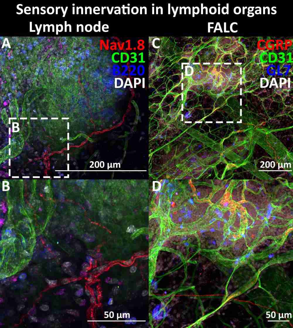
Figure 2: Confocal microscopy of sensory innervation in the lymph node and the omentum: Left (A,B): In lymph node tissue, Nav1.8 (red) reveals sensory nerve fibers within the subcapsular and vascular compartments in co-staining with CD31 (green) staining blood- and lymphatic vessles, B220 (blue) staining b cells, and DAPI (white) as a cell nuclei counterstain. Right: In fat-associated lymphoid clusters (FALC) of the omentum, CGRP (red) reveals sensory innervation in co-staining with CD31 (green) staining blood- and lymphatic vessles, GL7 (blue) staining germinal center b cells, and DAPI (white) as a cell nuclei counterstain. Insets (B,D) show zoomed image of the boxed areas above. Image: INI Research / Feline Bludau, Leonie Dührkoop.
- Neuronal regulation of FALCs: Leonie Dührkoop and Dr. Katja Jarick are investigating the innervation of fat-associated lymphoid clusters (FALCs) in the omentum under steady state and inflammatory conditions. Building on previous findings of increased innervation in activated FALCs, they systematically quantify sensory, sympathetic, and VIP+ neuronal input across different models, including S. ratti infection and inflammatory bowel disease. Their work aims to better understand how inflammation shapes neuro-immune interactions within the omental microenvironment.
- Patterns of innervation in three-dimensional lymph nodes using X-Clarity –alterations in inflamed lymph nodes: Merle Bannick is working on inflammatory models (S. ratti infection) to see whether or not there are differences in the Innervation pattern compared to steady state. The Tissue Clearing method allows a three-dimensional imaging of neuronal structures in the lymph node.
- Innervation of follicles and nodules in steady state and inflammatory models: Nenya Leising is working on an analysis of the innervation of nodules and follicles and directly comparing whether there is a difference in innervation pattern in the steady state vs. inflammatory model (S. ratti infection) and what this could mean for the nodule’s role in humoral immune responses during infection.
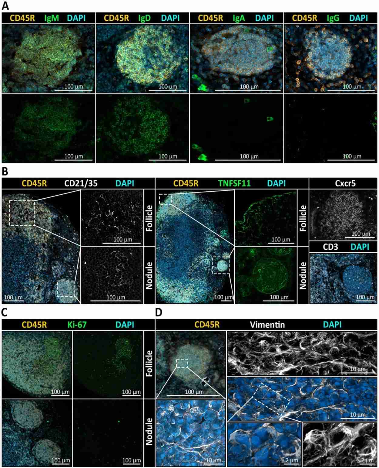
Figure 3: Stromal and chemokine-related markers suggest follicle-like organization of nodules. Left: Co-staining for CD45R (B cells), CD21/35 (follicular dendritic cell marker), and DAPI reveals a clear CD21/35+ FDC network in cortical follicles and in medullary nodules. Middle: TNFSF11 (RANKL), a marker of marginal reticular cells (MRCs), is detectable at the border of follicles as well as delineating nodules. Right: CXCR5, a chemokine receptor mediating B cell migration toward CXCL13-enriched regions, is detected in both follicles and nodules, while CD3+ T cells are infrequently observed and only sparsely distributed within nodules. Image: INI Research/ Paul Schütz.
Our Publications:
- Follicle-like niches outside the cortex? 3D phase-contrast µCT revealed medullary B cell nodules in mucosa-draining lymph nodes (2025):
https://www.frontiersin.org/journals/immunology/articles/10.3389/fimmu.2025.1674997/full
- Three-dimensional analyses of vascular network morphology in a murine lymph node by X-ray phase-contrast tomography with a 2D Talbot array (2022):
https://www.frontiersin.org/journals/immunology/articles/10.3389/fimmu.2022.947961/full
- GFAP and desmin expression in lymphatic tissues leads to difficulties in distinguishing between glial and stromal cells (2021):
https://www.nature.com/articles/s41598-021-92364-z
- Immunological memory: resting on two shoulders at antigen entrance sites? (2019):
https://www.nature.com/articles/s41423-019-0263-8
- Neural architecture in lymphoid organs: Hard‐wired antigen presenting cells and neurite networks in antigen entrance areas (2018):
https://pmc.ncbi.nlm.nih.gov/articles/PMC5946157/
- Dendritic cells and macrophages neurally hard-wired in the lymph node (2015):
Dendritic cells and macrophages neurally hard-wired in the lymph node | Scientific Reports
Our Team:
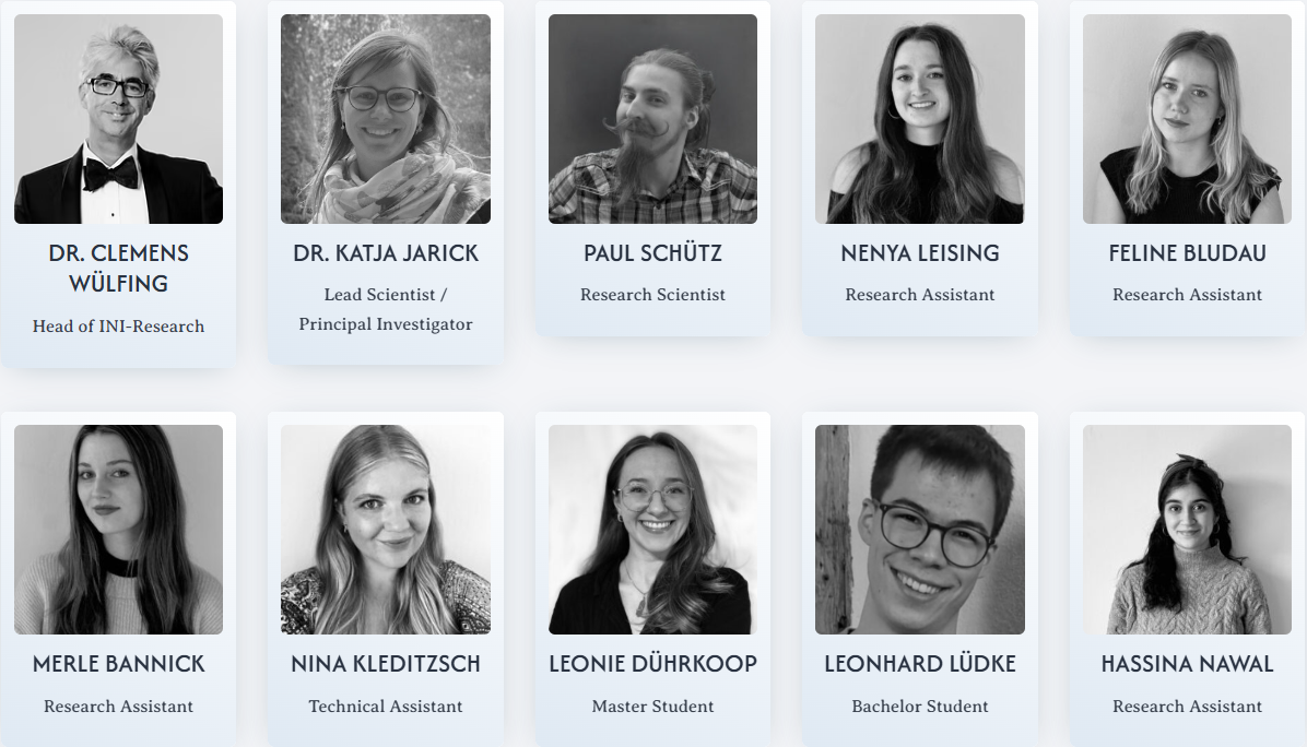
- Dr. Clemens Wülfing, lecturer of neuroimmunology and human biology, serves as the head of INI-Research.
- Dr. Katja Jarick leads scientific coordination within the group, supported by Nina Kleditzsch, our technical assistant.
- Paul Schütz, our research scientist, is responsible for bioinformatics and data analysis.
- Four Scholarship holders — Merle Bannick, Nenya Leising, Feline Bludau and Hassina Nawal — contribute to their own ongoing experimental and analytical projects.
- Master’s student Leonie Dührkoop and bachelor’s student Leonhard Lüdke, who are pursuing their thesis projects within the laboratory.
Related stories you may like:
The Nervous System’s Relationship to Immune Events: ‘Broadly Based but Cell-Faced’
Innervation of the Human Thymus and Spleen
Thymus Emergence & Evolution of Immune and Neuroendocrine Systems
Neural Meshwork Resembling Glass Fishing Float Envelops Antigen Presenting Cells in Lymphoid Organs
Aging Alters Sympathetic Noradrenergic Innervation and Immune Reactivity in the Lymphoid Organs: Strategies to Reverse Neuro-Immune Senescence